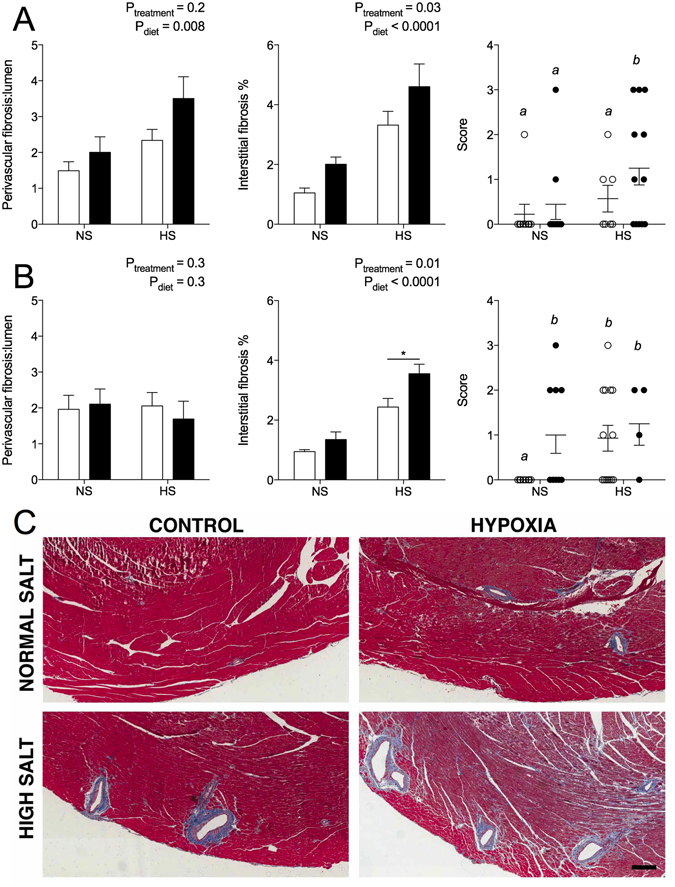
Figure 3.

Cardiac histopathology of offspring at 12 months of age. Perivascular fibrosis area normalised to lumen area, interstitial fibrosis expressed as percentage of cardiac tissue, and histology score in male (A) and female (B) offspring. (C) Masson’s Trichrome staining of cardiac tissue in males. Blue staining marks collagen (fibrosis). Scale bar represents 200 µm. Scoring analysed via one-way ANOVA with letters denoting statistical differences between groups. Perivascular and interstitial fibrosis analysed via two-way ANOVA. *P < 0.01 by Sidak post hoc. Values are mean ± SEM. Male: N = 5–11/group; female: N = 4–8/group. Control: open bars/points; hypoxia: closed bars/points.
