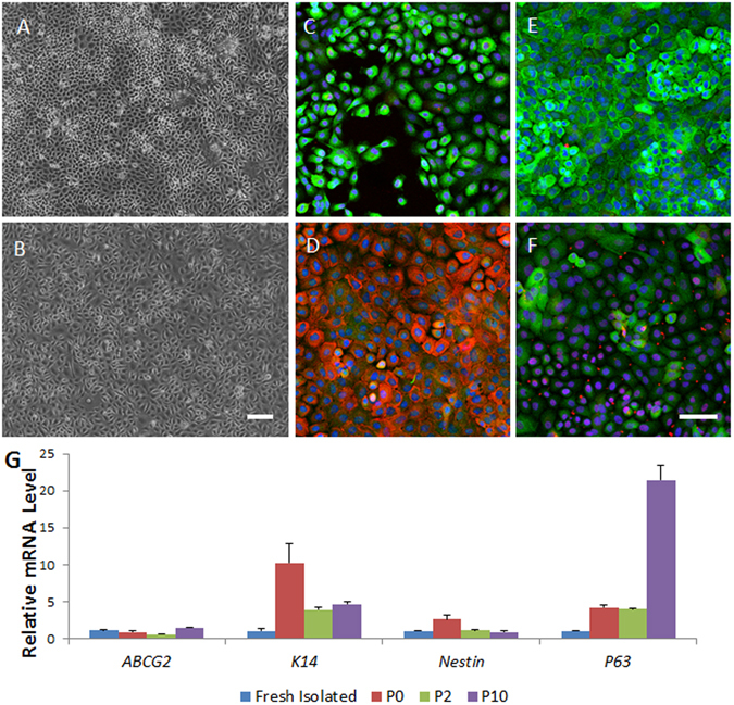
Figure 7.

The proliferative capability and characterization of the immature lacrimal gland epithelial cells in monolayer cultured in serum-free media. Monolayer culture of cells from day 7 samples after duct ligature release can form a compactly organized epithelial morphology from passage 0 (P0, A) to P12 (B). Immunostaining showed most of the cells to be nestin positive (green, C), ∆Np63 positive (red, C,F), ABCG2 positive (red, D), PCK positive (green, D) and K15 positive (green, E). Some of these P10 cells to be K14 positive (green, F), RT-qPCR showed the mRNA level of progenitor cell-related genes in the cultured cells (G). Bar represents 100 μm.
