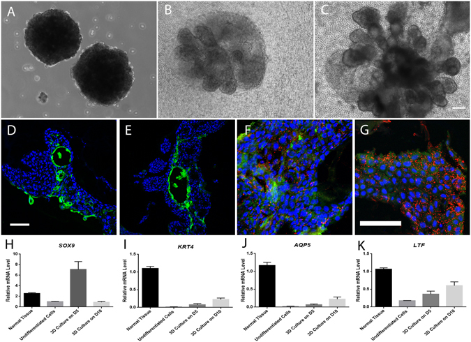
Figure 8.

3-D culture of lacrimal gland spheres. Lacrimal gland epithelial cells from day 7 post-reopening in P2 formed similarly sized lacrimal spheres in suspension culture (A), and gradually branched out in about 2 weeks (days in culture, 5, (B); 10, (C)) to form mini gland-like structure in laminin gel. Immunostaining demonstrated K4 (green, D,E) positive, Lactoferrin (red, F,G) positive and AQP5 positive (green, F,G) cells in those structures. RT-qPCR showed the mRNA level of differentiated lacrimal gland cell-related genes in the gland-like structures (H–J). Blue for DAPI. Bar represents 100 μm.
