Figure 6.

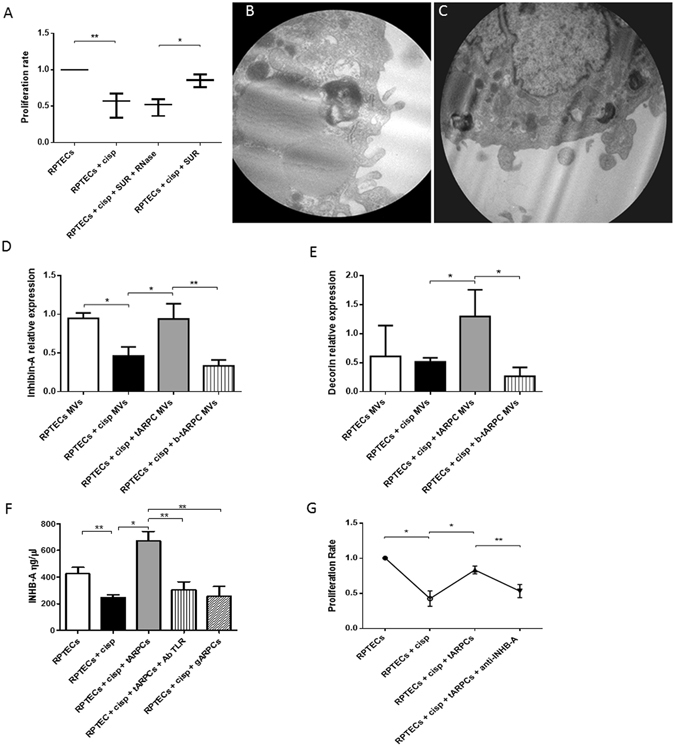
Inhibin-A and decorin were involved in the RPTEC repair. (A) Preconditioned supernatants from co-cultures induced an increase of RPTEC proliferation rate. Supernatants treated with 1 U/ml Rnase did not influence the proliferation rate. (B–C) Representative micrographs of transmission electron microscopy showing the release of MVs from the surface of a tARPC. Micrographs show the extrusion of MVs from the surface of the tARPC. Ultrathin sections, stained with led citrate were viewed by ZEISS EM910 electron microscope. Image acquisitions were performed with magnification of ×16000. (D) MVs isolated from the medium of regenerative condition (cisplatin-damaged RPTECs in co-culture with tARPCs), carried high levels of Inhb-A mRNA, these levels are comparable to the ones found in the supernatant of non-damaged RPTECs. Moreover, Inhb-A levels sharply decreased when TLR2 on tARPCs was blocked. (E) MVs isolated from the medium of the regenerative condition (cisplatin-damaged RPTECs in co-culture with tARPCs), carried high levels of Decorin mRNA, these levels were even higher than the ones detected in the supernatant of non-damaged RPTECs. Moreover, Decorin levels sharply decreased when TLR2 on tARPCs was blocked. (F) Inhb-A protein level increased in the regenerative condition (cisplatin-damaged RPTECs in co-culture with tARPCs), and this increase was abrogated when TLR2 receptor was blocked reaching levels similar to those obtained in the damaged condition. Inhb-A did not increased in the co-cultures of gARPCs with damaged-RPTECs. (G) BrdU cell proliferation assays showed that if we treated the regenerative medium (cisplatin-damaged RPTECs in co-culture with tARPCs) with the Inhb-A blocking antibody the damaged-RPTEC failed to recover their proliferation rate. Plots represent 5 independent experiments using tARPCs from 5 different subjects; *P < 0.05, **P < 0.005.
