
Fig. 2.
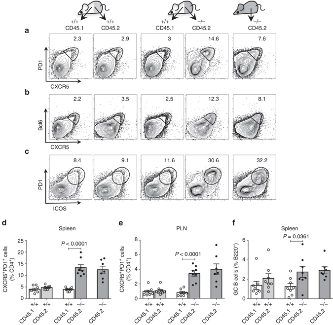
Expansion of TFH cells in DKO mice is cell-intrinsic. Analysis of mixed bone marrow chimeric mice (n = 8) 8–10 weeks after the reconstitution in Rag-1-deficient hosts. Chimeric mice were generated with 1:1 ratio of wild-type CD45.1 and wild-type CD45.2 bone marrow cells (control) or wild-type CD45.1+ and DKO CD45.2+ bone marrow cells (experimental) or only DKO CD45.2+ bone marrow cells (reference). a–c FACS plots for CXCR5 and PD1 a or CXCR5 and Bcl6 b or ICOS and PD1 c expression gated on CD4+ T cells. d, e Frequencies of TFH cells in the chimeric mice of the indicated genotypes. f Frequencies of GC B cells in the chimeric mice of the indicated genotypes. PLN peripheral lymph nodes. Each dot represents an individual mouse. Error bars indicate mean ± s.d., P-value by two-tailed t-test. Combined data from two independent experiments