
Figure 6.
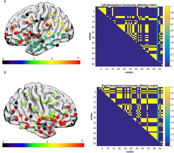
(A) Exemplar data Subject 2, each color represents a single community left hemisphere lateral view. Both hemispheres did not reveal dramatic fragmentation patterns. Subject 2 had a lesion volume of 76.1 cm3, percent white matter damage of 0.099, and a WAB-AQ score of 88.1. The community affiliation matrix showed relatively stable clustering over 100 runs. (B) Exemplar data Subject 2, right hemisphere lateral view, each color represents a single community, and again there was no apparent fragmentation, and the network was divided into 4 communities. The community affiliation matrix showed stable clustering over 100 runs.