
Figure 4.
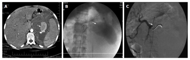
Splenic artery pseudoaneurysm before and after embolization. A: Post contrast computed tomography scan showing the pseudoaneurysm rising from the splenic artery; B: Pre embolization selective splenic arterial DSA angiography image showing pseudoaneurysm; C: Post embolization DSA image showing the coils inside the splenic artery with its resultant embolization[92].