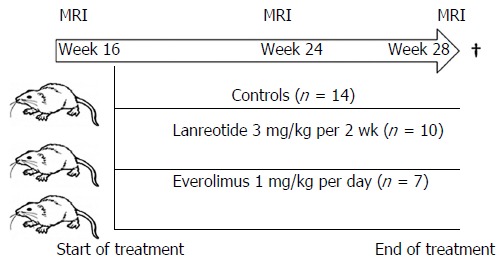
Figure 1.

Study design. Female PCK rats were obtained at age week 10. At week 16, a first MRI was performed to calculate the liver volume. Animals were randomly assigned to one of the groups and treatment was started. At week 24 and week 28 a new MRI was performed, after the last measurement the animals were sacrificed and tissue collected for protein and gene assay.
