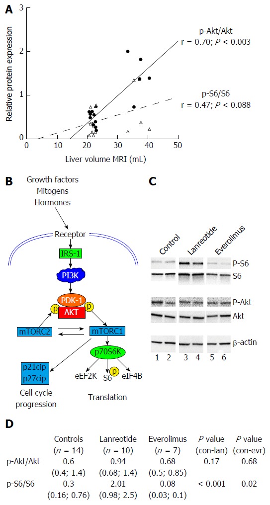
Figure 4.

Analysis of protein expression of components of PI3K/AKT/mTOR pathway by Western blot in livers of PCK rats after 12 wk of treatment. A: phosphorylation ratio in untreated animals: correlation between p-Akt/Akt ratio and p-S6/S6 ratio with liver volume. Pearson correlation (r) is given together with significance; B: Simplified schematic presentation of PI3K/AKT/mTOR signaling pathway; C: shows two representative samples from each experimental group; D: shows relative expression mean (range) of phosphorylated Akt vs total Akt (p-Akt/Akt) and S6 vs total S6 (p-S6/S6) with n as number of animals analyzed. Expression of proteins was normalized to β-actin levels.
