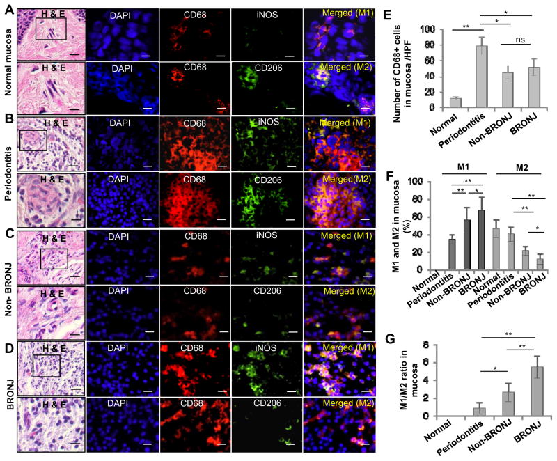
Figure 2.
Altered M1 and M2 macrophage infiltrations in oral mucosal tissues bordering the non-healing extraction socket of BRONJ patients. A–D, Immunofluorescence studies showed an increased infiltration of CD68+ iNOS+ M1 macrophages and a decreased infiltration of CD206+ M2 macrophages in mucosal tissues bordering the extraction sockets of patients manifested BRONJ lesions (BRONJ) or patients with history of zoledronate treatment without clinical BRONJ (Non-BRONJ) (n=5), whereby oral mucosal tissues from healthy patients who underwent routine dental extraction for other non-inflammatory mucosa conditions (Normal) or patients with diagnosis of inflammatory gum disease, specifically, active periodontitis with resultant tooth loss (Periodontitis) were used as controls (n=5). Scale bars, 50μm. E, Quantification of total CD68+ macrophages in 6 randomly selected high-power fields (HPFs). F and G, Quantification of M1 and M2 macrophages in 6 randomly selected high-power fields (HPFs). Data are mean ± SEM of multiple fields in n=5 per group. *P < 0.05; **P<0.01; ns, no significant statistical differences.

