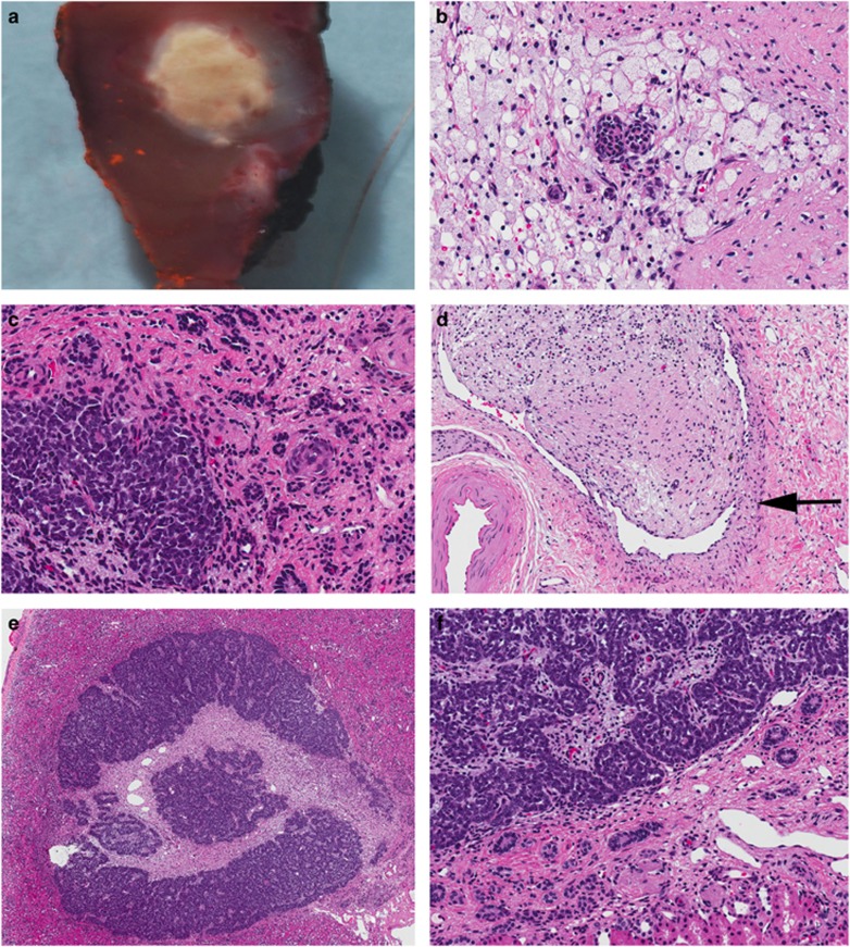
Figure 4.
Pathology findings from partial nephrectomy. (a–d) Treated WT. (a) Macroscopic appearance of the tumour measuring only 0.5 cm in diameter. (b) The central portion of the specimen is mainly necrotic with isolated differentiated epithelial elements. (c) The more peripheral portion consists of blastema (lower left corner) with differentiated tubules in a fibrotic stroma. (d) Intra-renal vein (vessel wall marked by arrow) containing a tumour thrombus composed of fibrous stroma and rare differentiated tubules. (e and f) perilobar nephrogenic rest showing a well circumscribed rest surrounded by normal renal parenchyma. The rest is composed of nests of tubules separated by varying amounts of fibrous stroma. (original magnifications: a × 1, b, c × 200, d × 100, e × 10, and f × 100).

