Figure 3.
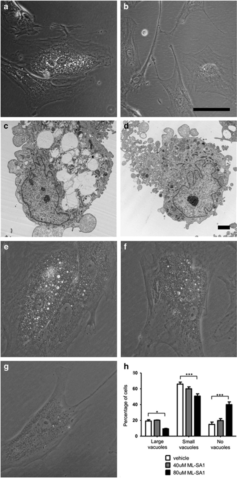
Prominent intracytoplasmic vacuoles were observed in a subset of patient-derived fibroblasts (a,c) but not in control fibroblasts (b,d). (a,b) phase-contrast microscopy (scale bar=100 μm). (c,d) electron microscopy (scale bar=2 μm): vacuoles have an ‘empty’ ultrastructural appearance and are delimited by a single membrane. Similar (less obvious) findings were also observed in positive control fibroblasts from a patient with FIG4-related YVS (data not shown). (e–h) Treatment of cells with a pharmacological TRPML1 activator (ML-SA1) resulted in dose-dependent rescue. Patient cells (treated with 0, 40, or 80 μmol/l ML-SA1 for 24 h) were scored as containing ‘large vacuoles’ (e), ‘small vacuoles’ (f), or ‘no vacuoles’ (g). >125 cells were scored for each condition in each of three biological replicates; two-way ANOVA (h) showed a significant (*P<0.05; ***P<0.001) increase in cells with ‘no vacuoles’ after treatment with 80 μmol/l ML-SA1.

