
Figure 22.
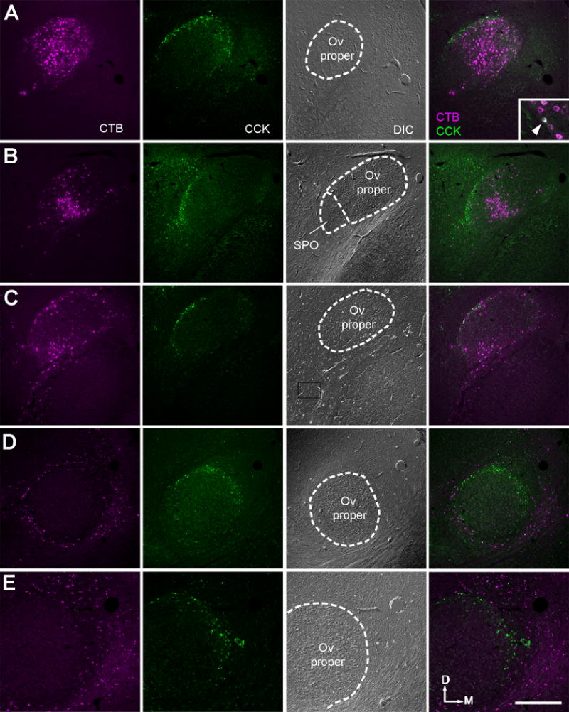
Retrogradely labeled neurons in the Ov complex and surrounding area following CTB injections into forebrain areas. The three left columns are single channel confocal images of CTB, immunoreactivity for CCK, and DIC, respectively. The far right column is the merged images of CTB and CCK. A–C, Retrogradely labeled neurons in Ov are located inside of or overlapped with the cOv indicated by CCK-immunoreactive cells, following injections into the Field L. Double-labeled cells were occasionally seen (Insert in A; arrowhead). D, Retrogradely labeled neurons are located outside of the cOv indicated by CCK-immunoreactive cells, following injections into the nucleus taenia of the arcopallium. E, Higher magnification images of the same case in D. The location is comparable to the box in D. No double-labeled cells were found. The dorsal is up and the right is medial. Abbreviations: see table of abbreviations. Scale bar = 500 μm.