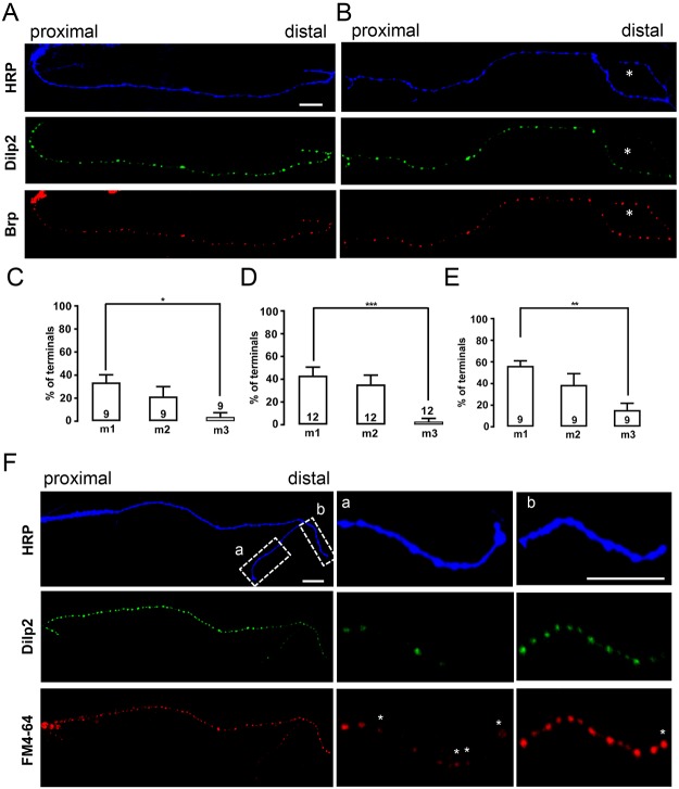
Fig. 1.
Neuropeptide stores, active zones and endocytosis in distal type II boutons. (A) Neuronal membrane (HRP), Dilp2–GFP and Brp in type II boutons on muscle m1. All images are oriented with distal boutons on the right. Dilp2–GFP expression was driven by Tdc2–Gal4 and terminals are labeled by TRITC–HRP immunofluorescence. In this example, DCVs and active zones are widely distributed in boutons. (B) Similar experiment to that in A, except that the most-distal boutons (marked with asterisks) contain less Dilp2–GFP, although Brp is present. (C–E) Quantification of the percentage of axonal branches on the indicated muscles in Tdc2>Dilp2-GFP animals (C), Tdc2>ANF-GFP animals (D) or elav>ANF-GFP animals (E) lacking neuropeptide content in distal boutons.The number of examined neurons, each from an independent animal is indicated for each muscle. For each neuron, all the branches on each muscle were imaged and the number of branches with dimmer distal boutons was divided by the total number of branches to calculate the percentage for each muscle. One-way ANOVA with Tukey's post-test was performed to determine statistical significance. *P<0.05, **P<0.01, ***P<0.001. (F) Comparison of Dilp2–GFP and uptake of the SSV marker FM4-64 on m2. Asterisks indicate boutons with FM4-64 that lack Dilp2–GFP. Scale bars: 10 µm.

