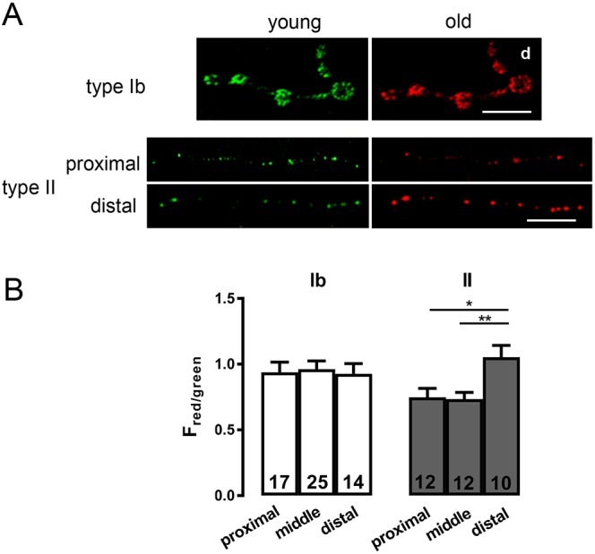
Fig. 3.

DCV age differs in proximal and distal boutons. (A) Age-dependent labeling of DCVs containing ANF tagged with the mK-GO timer protein driven by 386Y-Gal4 in type Ib and type II boutons on m2. The green (young) and red (old) images are shown side by side. Scale bars: 10 µm. For type Ib image, the distal boutons are on the right (labeled d), while separate images are shown for proximal and distal regions containing type II boutons. (B) The ratio of red to green fluorescence in type Ib and type II boutons. Number of terminals analyzed in 12 animals is indicated for each region and bouton type. One-way ANOVA and Tukey's post-tests were performed to determine statistical significance of differences between red to green ratios in proximal, middle and distal regions containing 7 to 8 boutons each. **P<0.01, *P<0.05.
