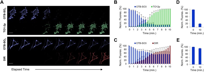
Fig. 4.
Determining fluorescence signal exchange at the surface of live cell via two-fluorogen competition. All analyses were performed using mammalian cells expressing HL1.0.1-TO1 fused to ADRB2 at the cell surface. (A) Time-lapse micrographs from cells initially labeled with OTB-SO3 fluorogen, then presented in the medium with either TO1-2p or DIR fluorogens and immediately time-lapse imaged for 10 min. Note, we observed improved image resolutions at longer wavelength emissions due to reduced cellular background and higher laser excitation power settings. Each fluorogen time-series micrograph represents multiple images over time. Scale bar: 15 μm. (B) Bar graph summary of fluorescence intensities from fluorogen competition time-lapse images of OTB-SO3 vs TO1-2p. The TO1-2p signal was normalized to the fluorescence values for the final time point. (C) Bar graph summary of fluorescence intensities from fluorogen competition time-lapse images of OTB-SO3 vs. DIR. The DIR signal was normalized to the fluorescence values for the final time point. (D) Bar graph summary of OTB-SO3 fluorescence at only two time points after addition of TO1-2p in the medium. (E) Bar graph summary of OTB-SO3 fluorescence at only two time points after addition of DIR in the medium. All assays were performed in presence of 100 nM of each fluorogen, and each bar graph analysis was determined from n=5 images for each group. Results are mean±s.d.

