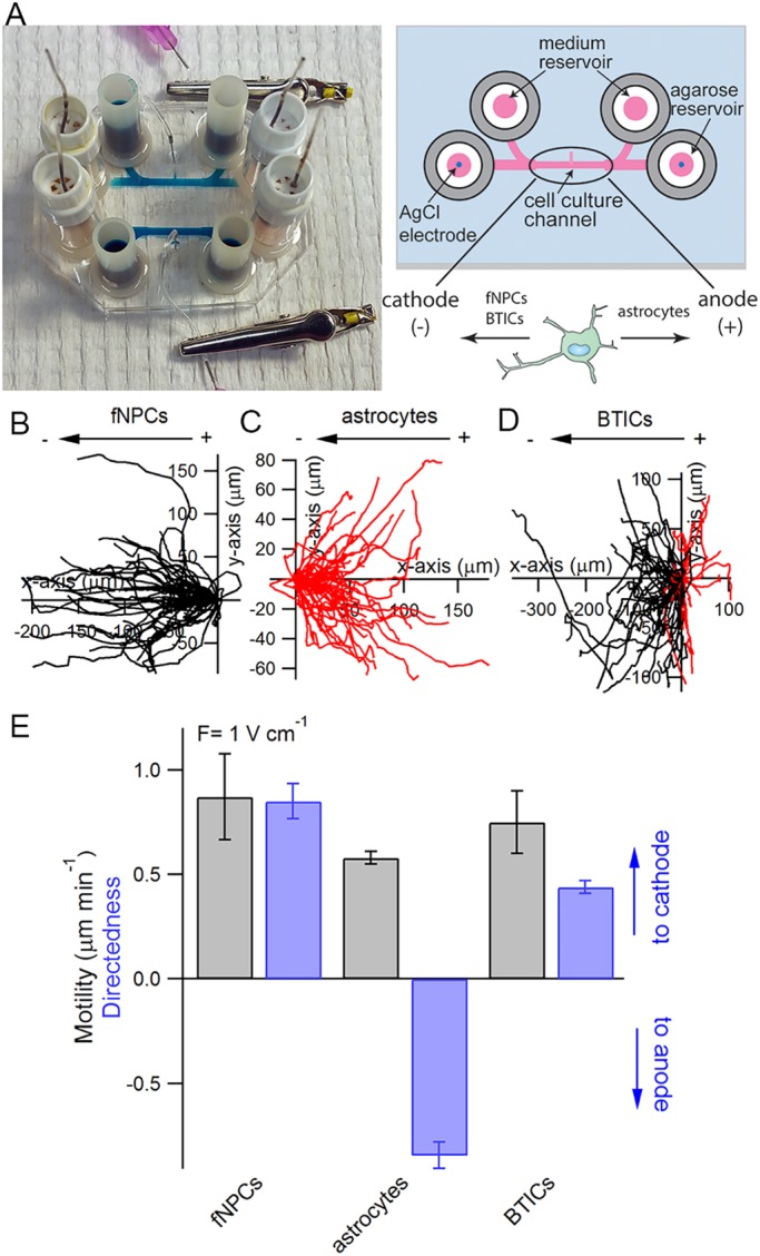
Fig. 1.

Galvanotaxis of fetal neural progenitor cells (fNPCs), astrocytes and brain tumor-initiating cells (BTICs). (A) Each galvanotaxis chip contains two symmetrical devices on a 35 mm×50 mm glass coverslip. Each device features two coiled Ag/AgCl electrodes embedded in agarose reservoirs located at each end of the cell culture channel, along with two media reservoirs also located at each end of the channels. The dimensions of the cell culture channel are 10 mm×1 mm×250 μm (length×width×height). A cell injection port located in the middle of the cell culture channel is used to introduce cells into the device and is clamped with an alligator clip afterwards to prevent evaporation. Trajectories of fNPCs (B), astrocytes (C) and BTICs (D) in the presence of an EF are analyzed and overlaid at the origin to characterize the galvanotaxis of each cell line. Each trajectory represents the actual path traveled by a cell in 3 h either to the cathode (left, black) or anode (right, red). (E) Quantitative analysis of cell motility and directedness in the presence of a 1 V cm−1 EF.
