
Figure 4. Pix and Git function as a bipartite scaffold to dimerize and activate Hippo.
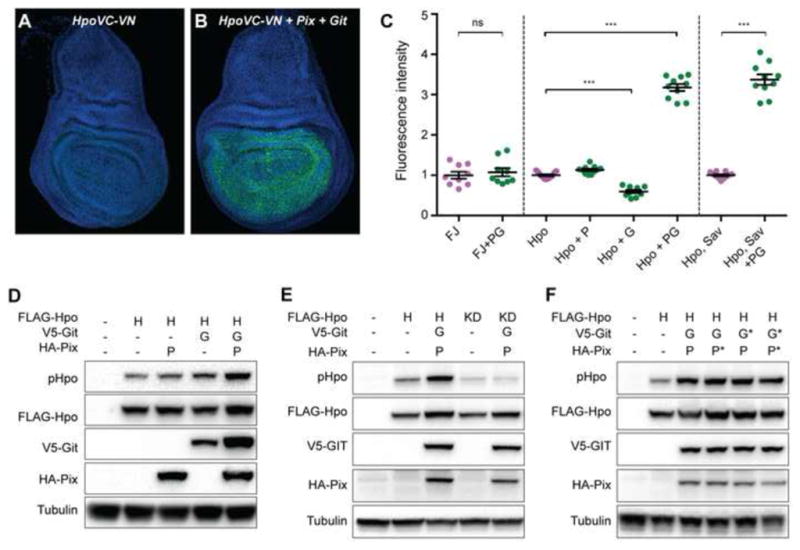
(A and B) Third instar larval wing imaginal discs stained with DAPI (blue). Both tissues express Hpo kinase-dead proteins fused to either the N- or C-terminus of Venus fluorescent protein (green). The tissue in (B) also expresses Pix and Git. (C) Quantification of Venus fluorescence in wing larval wing imaginal discs of the indicated genotypes (F=Fos, J=Jun, P=Pix, G=Git). n=9, 9, 11, 11, 10, 10, 11 and 10 from left to right. Data represents mean +/− SEM. *** indicates a p-value <0.001, whilst ns indicates no significant difference. (D–F) Western blot analysis of protein lysates from S2 cells transfected with the indicated plasmids. Hpo phosphorylation was detected using an antibody recognising phosphorylation of the Hpo activation loop (T195). Western blots were also probed with Anti-Flag, Anti-HA, Anti-V5 and Anti Tubulin to reveal total Hpo, Pix, Git and tubulin respectively. See also Figure S4.