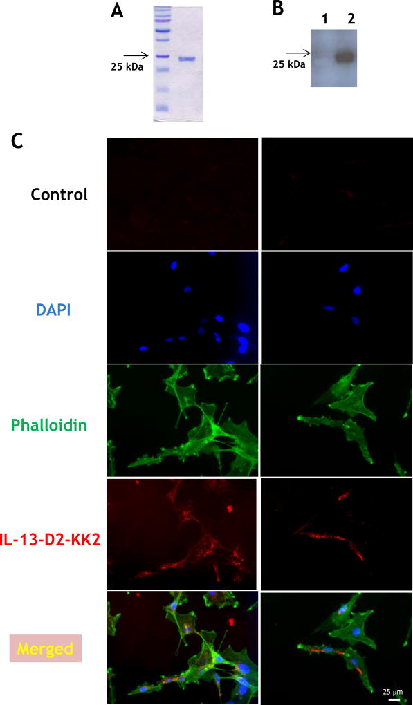
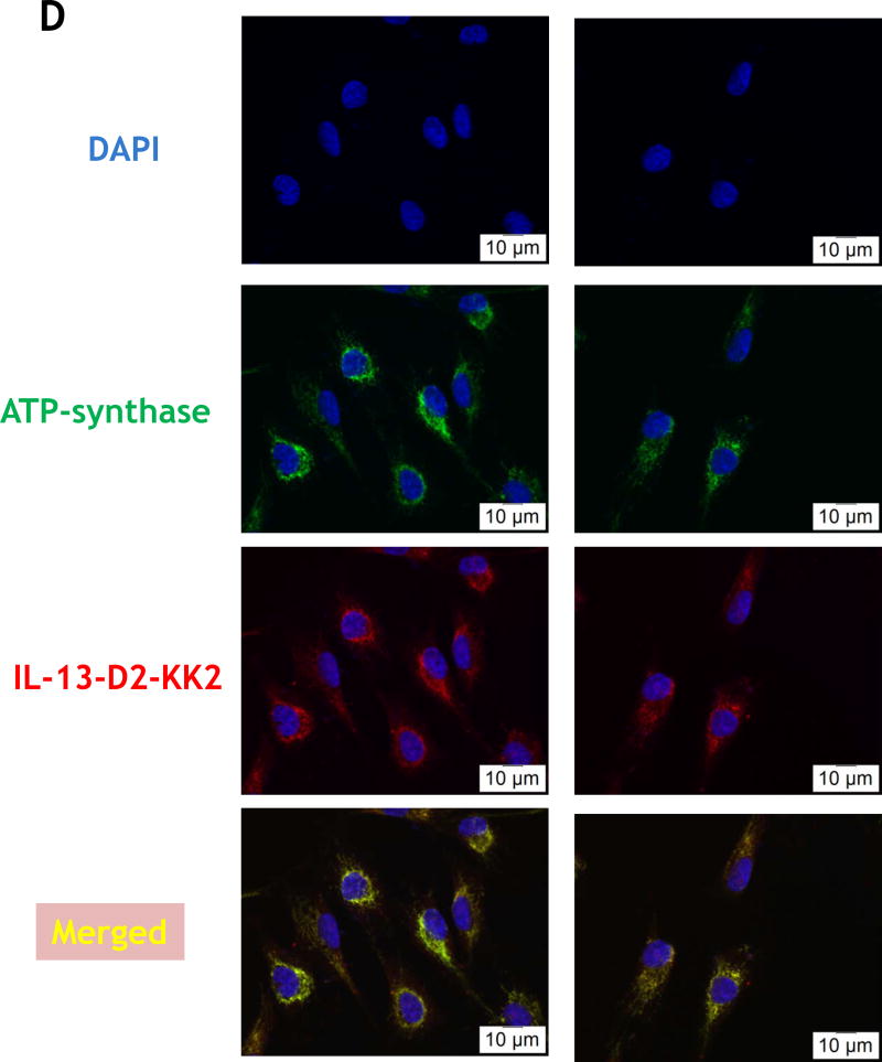
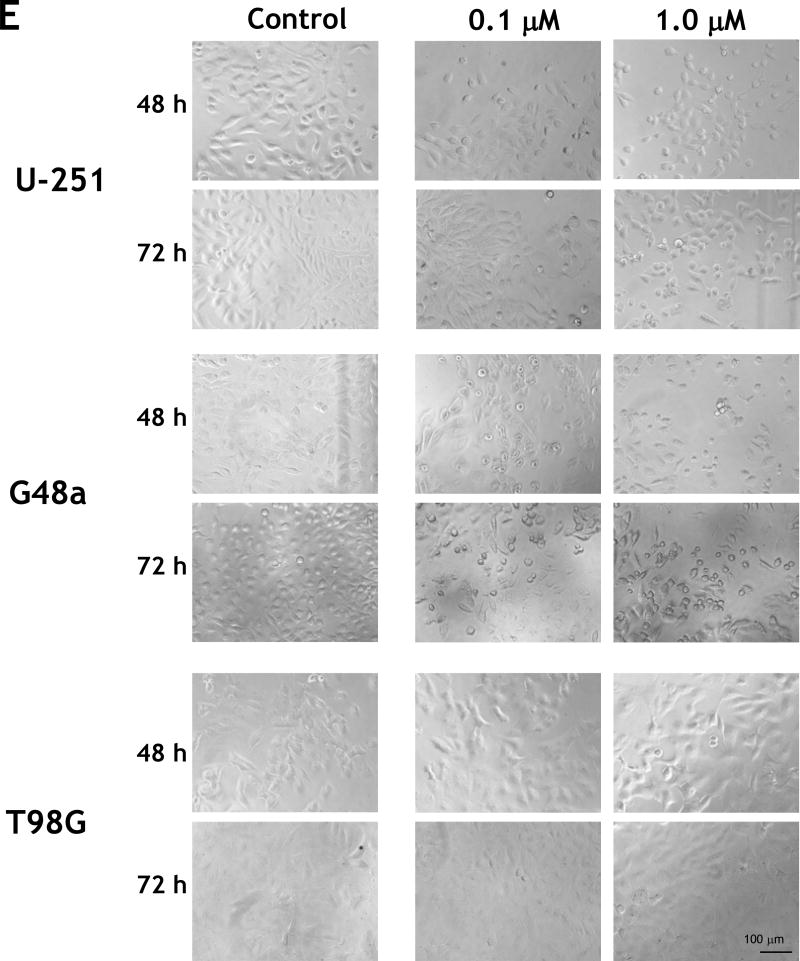
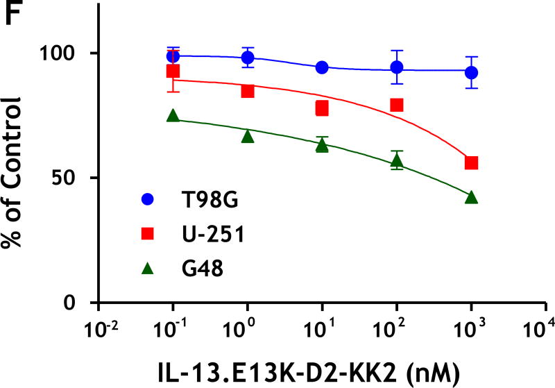
Figure 3.
(A) Purified recombinant IL-13-D2-KK2 protein. SDS-PAGE stained with Coommassie blue. (B) Immunoblot of biotinylated IL-13-D2-KK2 probed with Streptavidin-HRP. (C) Cell internalization of biotinylated IL-13-D2-KK2. The protein was analyzed by using anti-streptavidin Alexa Fluor 555 and fluorescent microscopy. (D) U-251 cells were treated with biotinylated IL-13-D2-KK2 for 8 hrs and the co-localization of biotinylated IL-13-D2-KK2 with mitochondrial ATP-synthase enzyme was analyzed by confocal microscopy. (E) IL-13-D2-KK2 effect on GBM cell lines U-251 MG, G48a and T98G analyzed using phase contrast microscopy. (F) GBM cells were treated with IL-13-D2-KK2 for 72 hrs and the cytotoxicity was measured by an MTS assay.


