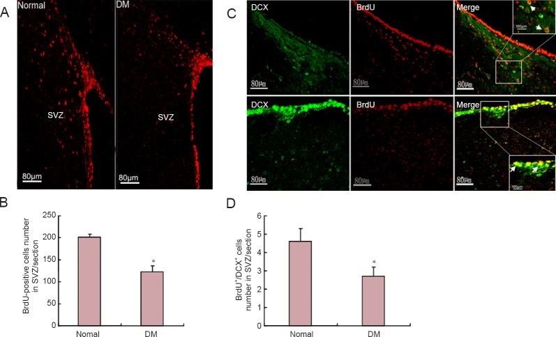
Figure 4.
Effect of DM on neural stem cell status in SVZ.
Representative images of BrdU staining and immunofluorescence histochemistry were observed by fluorescence microscope. (B) BrdU-positive cells were counted. Positive cell number decreased in the DM group compared with the normal group. (C) Representative images of BrdU and DCX double-labeling, with immunofluorescence staining observed by confocal microscopy. Arrows indicate double-labeled cells. Squares in right images are magnified in upper/lower corners. (D) BrdU+/DCX+ cells decreased in the DM group compared with the normal group. DM group: Overnight-fasted rats were injected once with 65 mg/kg streptozotocin through the femoral vein to induce DM. Normal group: Age-matched normal rats received an equivalent volume of normal saline. *P < 0.05, vs. normal group (mean ± SEM, n = 8). One-way analysis of variance was performed for multiple-group comparisons, with Tukey's post-hoc analysis performed for unpaired group comparisons. Experiments were performed in triplicate. BrdU: Bromodeoxyuridine; DCX: doublecortin; DM: diabetes mellitus; SVZ: subventricular zone.

