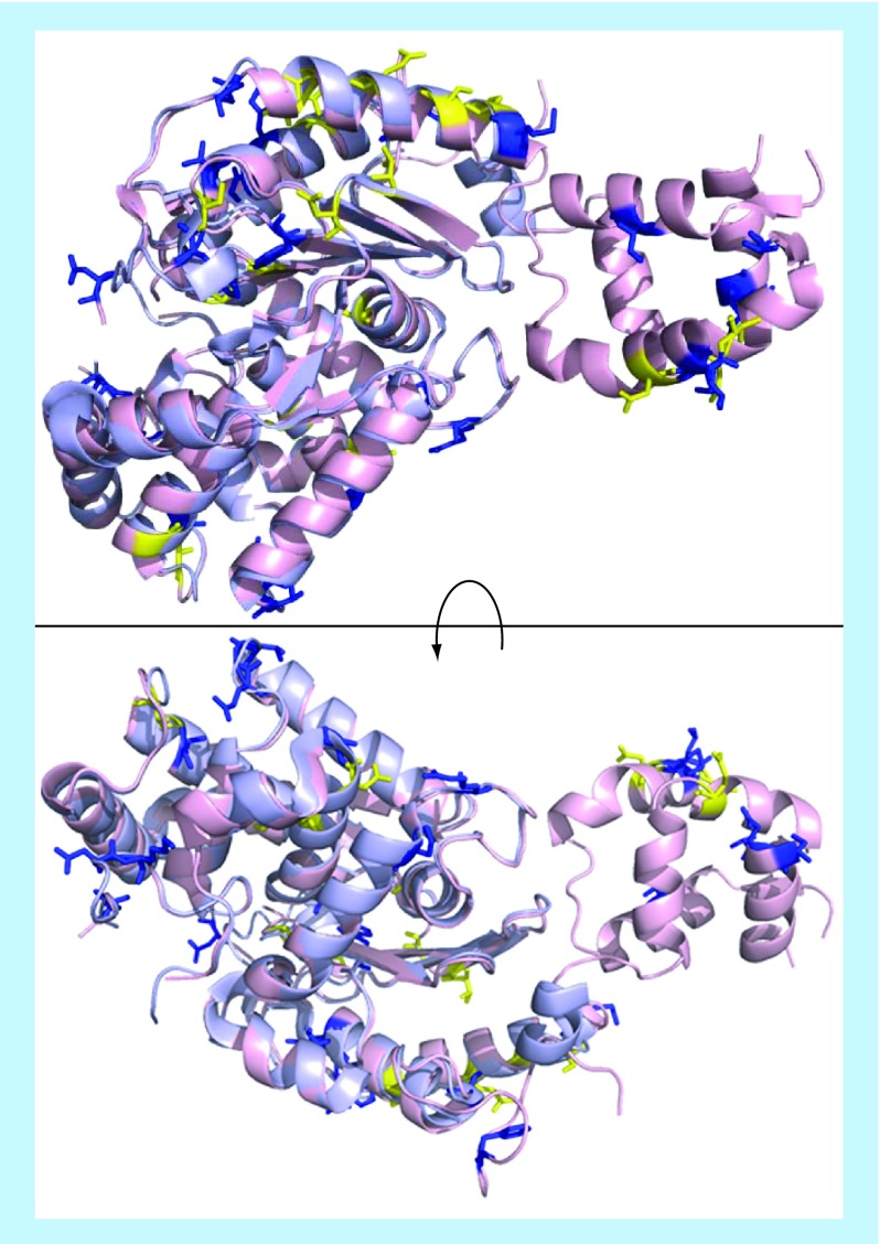
Figure 4. . Glutaminase C (light pink, crystal structure 5D3O) and liver-type glutaminase (light blue, crystal structure 4BQM) were aligned in PyMol.
Residues which are weakly similar or entirely dissimilar (Clustal alignment score of “.” or “ “, Figure 3) are drawn as sticks and shown in yellow and blue, respectively.

