Figure 2.
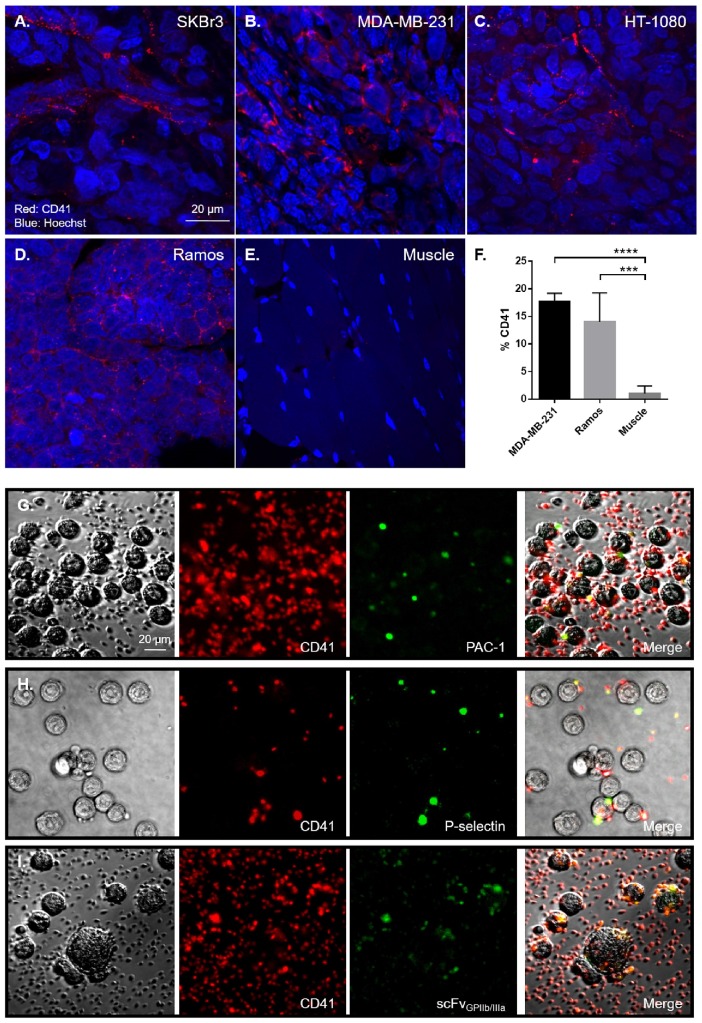
Platelets are abundant in human xenografts and are activated by tumour cells. Formalin fixed sections from human xenografts underwent immunofluorescence for platelets (red) and nuclei (blue) with an anti-CD41 antibody and counterstained with Hoechst® nucleic acid stain. Representative 60x magnification confocal fluorescence microscopy images of mice bearing SKBr3 (n=4), MDA-MB-231 (n=4), HT-1080 (n=4) and Ramos (n=4) tumour xenografts sections shows CD41 positive (red) staining (A-D), demonstrating that similar to human tumours, platelets are present within distinct mouse xenograft tumour tissues, compared to healthy muscles (E). Quantitative analysis of the number of platelets within MDA-MB-231 (n=3) and Ramos (n=5) tumours was performed by CD41 staining in flow cytometry, compared to healthy muscles (n=5) (F). To demonstrate the ability of tumour cells to directly activate platelets, SKBr3 cells were incubated with human platelets for 2 hours in vitro. Platelets were then stained with a CD41 antibody (red) and the platelet activation markers PAC-1 binding (green) (G), P-selectin expression (green) (H) and scFvGPIIb/IIIa binding (green) (I) was determined, demonstrating the presence of activated platelets on the tumour cells. Statistical analysis was performed using one way ANOVA and statistical significance was assigned for p values <0.001, represented by ***.

