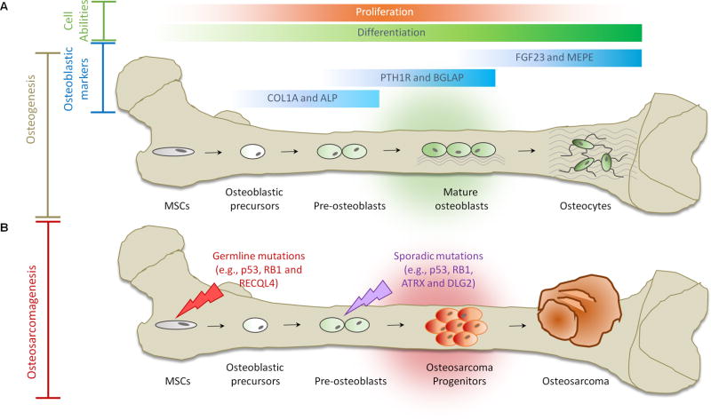
Figure 1. Osteogenesis and Osteosarcomagenesis.
(A) Initiation of osteogenic differentiation from MSCs. MSCs are multipotent bone marrow cells that are capable of differentiating to bone (osteoblast/osteocyte), fat (adipocyte) and cartilage (chondrocyte) tissues. Osteogenic differentiation is a tightly regulated process involving various signal transduction pathways (e.g., BMP and WNT), transcriptional regulators (e.g., p53, ZEB1, RUNX2 and ZNF521) and cell cycle controllers (e.g., RB1). Gene expression continuously changes through distinct osteogenic differentiation stages. COL1A and ALP are markers for osteoblastic progenitors and preosteoblasts. PTH1R and BGLAP serve as markers for mature osteoblasts. FGF23 and MEPE are markers for osteocytes. (B) Defects in osteogenesis lead to osteosarcomagenesis. Genetic alterations (e.g., germline mutations in p53, RB1 and RECQL4) probably interfere with the normal osteogenic process, resulting in incompletely differentiated osteoblasts or osteocytes in bone. These defects disrupt the balance between proliferation and differentiation and may cause a group of cells to display uncontrolled cell proliferation. Osteosarcoma progenitors may arise from these cells and expand to form osteosarcoma.

