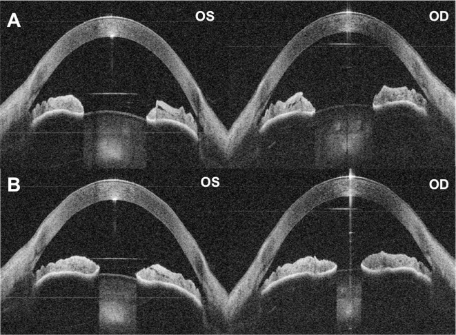
Figure 3.
Automated quantitative pupillometry using the binocular OCT prototype. (A) Pupils of both eyes dilated immediately prior to stimulation. (B) Pupils at maximum constriction after controlled flash of light presented to the right eye. Note the anisocoria – the left pupil does not constrict equally to the right pupil. In post-processing, the presence and extent of pupillary defects can be calculated using OCT-derived measurements of the pupil circumference.

