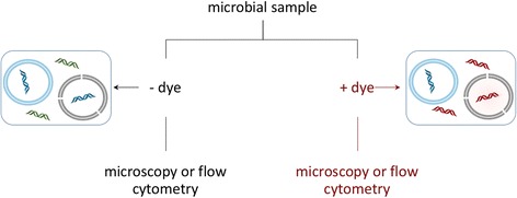
Fig. 3.

Live/dead staining workflow, propidium iodide (PI) example. In this technique, the sample is divided in two. One sample (left side) is stained with a total nucleic acid stain and used for cell enumeration, in which the live (blue membrane) and dead (black membrane) cells cannot be distinguished from each other, resulting in a stain of all nucleic acids. In the propidium iodide (PI) stained sample, the stain permeates compromised cell membranes, staining both cells presumed to be dead or in the process of dying (black membrane) and extracellular DNA or DNA, with PI-stained DNA colored red. Live cells with intact membranes (blue membrane) are not stained. In both types of samples, localization of stains within cells allows for enumeration, with stained free DNA relegated to background fluorescence. A comparison of counts from stained and unstained samples can be used to estimate the number of living cells. Alternatively, a single sample can be prepared with both a total nucleic acid stain and propidium iodide for counts of living and dead cells in the same preparation (not shown)
