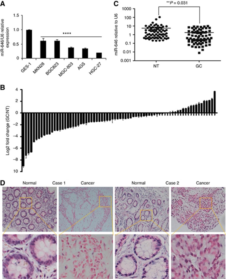
Figure 1.
miR-646 expression is downregulated in GC cell lines and tissues. (A) Decreased miR-646 expression was detected in all five GC cell lines compared with the normal human gastric epithelial cell line GES-1. ****P<0.001. (B) Real-time PCR analysis of miR-646 expression in 74 pairs of human GC tissues and their adjacent normal mucosal tissues. The error bars represent the mean±s.d. from three independent experiments. (C) As analysed by qRT–PCR, miR-646 expression level in GC tissues was significantly lower than that observed in the corresponding noncancerous gastric mucosa tissue. **P<0.05 (D) Representative ISH images of miR-646 expression. Scale bars, 50 μm.

