Figure 1.
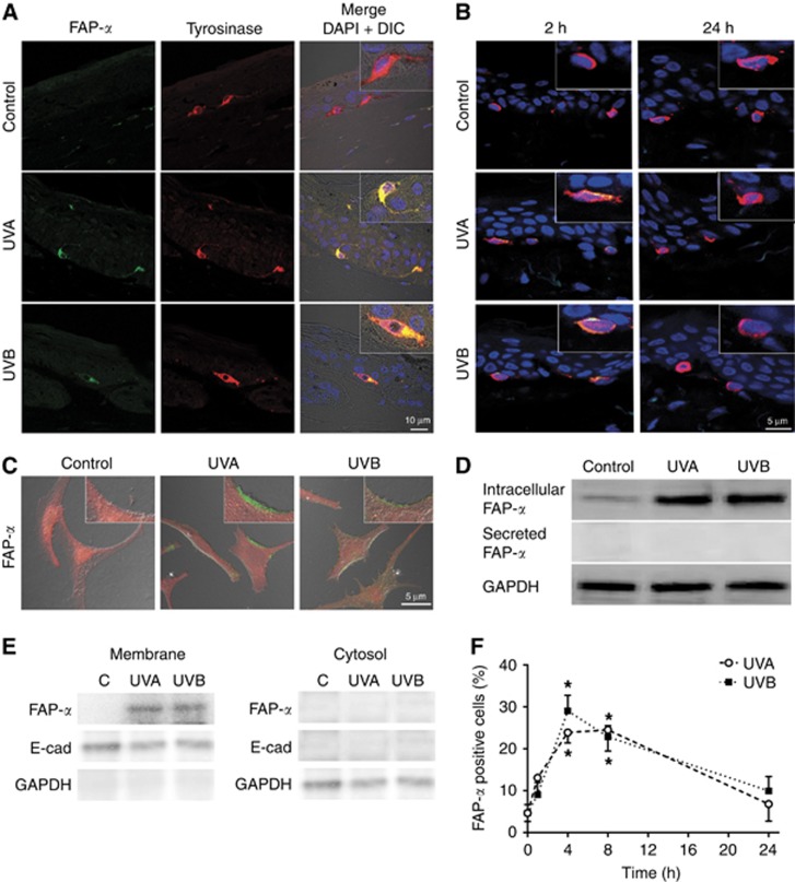
UV radiation augments FAP-α expression. Samples were irradiated with UVA (6 J cm−2) or UVB (60 mJ cm−2). (A) Artificial skin constructs stained for FAP-α (green) and the melanocytic marker tyrosinase (red) in combination with DAPI-stained nuclei (blue) and bright field in the merged image 4 h after UVR. (B) Normal ex vivo skin stained 2 and 24 h after radiation showing representative merged images of FAP-α (green), the melanocytic marker tyrosinase (red) and DAPI-stained nuclei (blue). (C) Representative merged images of immunofluorescence of FAP-α (green), cytosolic marker protein (LDH, red) and bright field, and (D) immunoblotting of active and secreted FAP-α 4 h after UVR. GAPDH is used as loading control. (E) Immunoblotting of FAP-α, E-cadherin (E-cad, plasma membrane marker), and GAPDH (cytosolic marker) in subcellular fractions after radiation. (F) Quantification of FAP-α-positive melanocytes following radiation (mean±s.d.; n=4; *P<0.05).

