Figure 3.
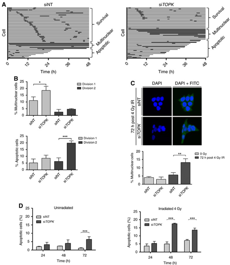
TOPK depletion causes multinucleation and apoptosis in HCT116 cells.Cells were irradiated 72 h post siRNA treatment. (A) Live-cell imaging following the fate of 50 HCT116 H2B-mCherry cells for 48 h after 4 Gy IR. Each horizontal bar represents one cell, with interphase shown in grey and mitosis shown in black. Following division, the first daughter cell to undergo division, apoptosis or multinucleation is shown. (B) The percentage of cells that apoptosed or became multinuclear during the first or second cell divisions after IR (n=3, average of 3 replicates). (C) Analysis of IR-induced multinucleation in fixed HCT116 treated cells 72 h after 4 Gy IR. At least 100 cells were counted per condition. Nuclei were stained with the DAPI DNA stain (blue) and FITC was used as a cytoplasmic counterstain (green). (D) Cell death in the presence (right panel) and absence (left panel) of 4 Gy irradiation was assessed with Hoechst staining to identify bright apoptotic nuclei in cells transfected with NT or TOPK siRNA and irradiated with 4 Gy at T=0. Data displayed are representative of three independent experiments and were analysed using unpaired two-sided t-tests; *P<0.05, **P<0.01 and ***P<0.001.

