Fig. 2.

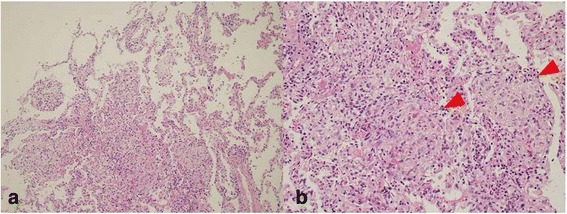
TBLB specimen from the right middle lobe showed intraalveolar granulation tissue with myofibroblasts consistent with organizing pneumonia (Hematoxin-Eosin (HE) × 100) (a). Masson body (red arrow) was seen on the TBLB specimen (HE ×400) (b)

TBLB specimen from the right middle lobe showed intraalveolar granulation tissue with myofibroblasts consistent with organizing pneumonia (Hematoxin-Eosin (HE) × 100) (a). Masson body (red arrow) was seen on the TBLB specimen (HE ×400) (b)