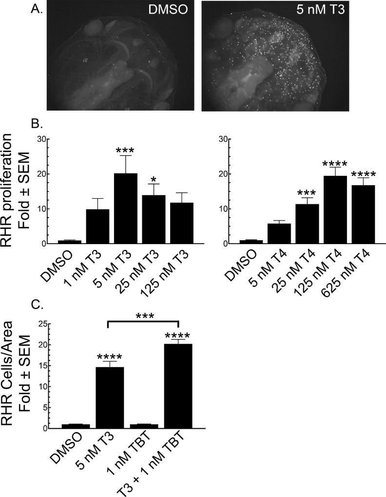
Figure 3.
T3 and T4 increase cellular proliferation in the rostral head area (RHR). A. Representative photomicrographs comparing the extent of phospho-Ser10 H3 staining in tadpoles treated for 5 days with vehicle (DMSO) or 5 nM T3. B. Quantification of RHR proliferation over a T3 (left panel) or T4 (right panel) concentration range normalized to vehicle (DMSO) control. C. The potentiation of RHR proliferation by co-treatment of 5 nM T3 with 1 nM TBT for four days and normalized to the resulting RHR area. Bars represent the mean with standard error (n = 15). Significantly different from vehicle control at *, p < 0.05; ***, p<0.001; ****, p<0.0001 as determined by 1-way ANOVA with Dunnett’s (for T3 or T4 alone) or Sidak’s (for T3-TBT) multiple comparison test (MCT).

