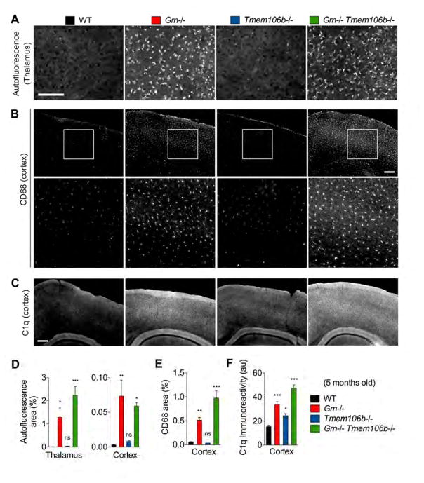
Figure 7. TMEM106B deficiency does not revert accumulation of lipofuscin, CD68-positive microglia, and complement C1q in Grn−/− mice.
(A) Representative images of autofluorescence using 488 nm excitation in thalamus of 5-month-old WT, Grn−/−, Tmem106b−/−, and Grn−/− Tmem106b−/− mice. Bar, 50 μm
(B) Representative images of WT, Grn−/−, Tmem106b−/−, and Grn−/− Tmem106b−/− cortex stained for CD68 at 5 months of age. Bottom panels are high-magnification of white square area in top panels. Bar, 200 μm.
(C) Representative images of WT, Grn−/−, Tmem106b−/−, and Grn−/− Tmem106b−/− cortex stained for C1q at 5 months of age. Bar, 200 μm.
(D) Quantification of autofluorescent puncta area (%) in 5-month-old WT, Grn−/−, Tmem106b−/−, Grn−/− Tmem106b−/− mice. Mean ± sem, n = 4–5/group, *p < 0.05, **p < 0.01, ***p < 0.001; One-way ANOVA with Dunnett’s post hoc test.
(E) Quantification of C1q-immunoreactivity in WT, Grn−/−, Tmem106b−/−, and Grn−/− Tmem106b−/− cortex. Mean ± sem, n = 3–5/group. *p < 0.05, ***p < 0.001; One-way ANOVA with Dunnett’s post hoc test.
(F) Quantification of CD68-immunoreactive area (%) in WT, Grn−/−, Tmem106b−/−, and Grn−/− Tmem106b−/− cortex. Mean ± sem, n = 3–5/group. *p < 0.05, ***p < 0.001; One-way ANOVA with Dunnett’s post hoc test.

