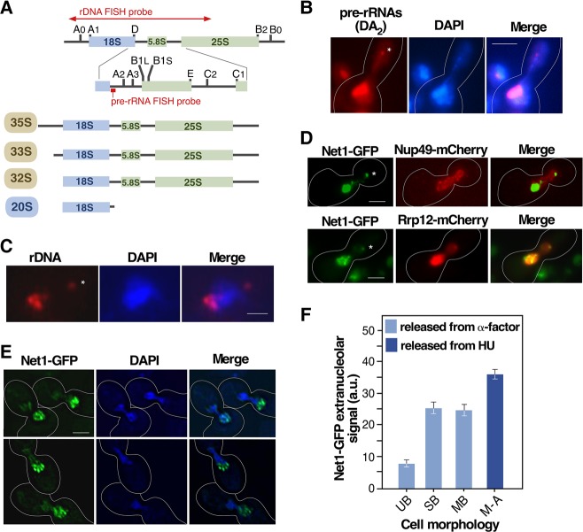
FIGURE 3.
The extranucleolar preribosome body contains both pre-rRNA and rDNA. (A) Scheme of the structure of the 35S pre-rRNA and intermediate rRNA precursors detected in the RNA FISH analyses shown in panel B. The position of the ITS-1 pre-rRNA probe is indicated. The 35S, 33S, and 32S pre-rRNAs are present in 90S particles, and the 20S pre-rRNA is present both in nucleolar and cytoplasmic pre-40S particles (see preribosome maturation pathway in Fig. 2A). The region encompassed by the rDNA FISH probe used for the experiment shown in panel C is indicated. (B) Representative image of the extranucleolar localization (indicated by asterisk) of pre-rRNAs in esp1-1 cells arrested in metaphase–anaphase that were analyzed by RNA FISH using a probe for the ITS-1 region. (C) Representative image of rDNA localization in esp1-1 cells arrested in metaphase–anaphase that were analyzed by DNA FISH. (D) Representative images of the subcellular localization of indicated GFP- (green color) and mCherry (red color)-tagged pairs of proteins in cells transiting metaphase–anaphase 40 min upon release from hydroxyurea arrest. (E) Representative images of net1-GFP-expressing cells transiting early mitosis upon release from α-factor arrest (top panels), and of esp1-1/net1-GFP cells arrested in metaphase–anaphase (bottom panels) taken under slow-bleach low-resolution conditions. (F) Quantification of Net1-GFP extranucleolar fluorescence in unbudded (UB), small bud (SB, bud size < 0.3× mother cell diameter), and medium bud (MB, bud size 0.3–0.6× mother cell diameter) wild-type cells released from α-factor arrest and wild-type cells at metaphase–anaphase (M–A) upon release from hydroxyurea arrest. HU, hydroxyurea. Scale bars (A–C), 2 µm.

