FIGURE 6.
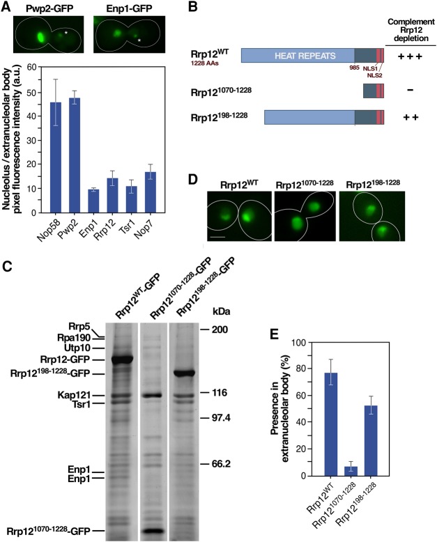
Protein localization at the extranucleolar body is a bona fide indicator of entry in the ribosome maturation pathway. (A) (Top) Representative images of the localization of indicated GFP-tagged proteins in the nucleolus and extranucleolar body (indicated by an asterisk). (Bottom) Quantification of the average fluorescence intensity per pixel detected for the indicated GFP-tagged proteins (x-axis) in the nucleolus and extranucleolar body. (B) Summary of the capacities of wild type and amino-terminal deleted versions of Rrp12 to complement the loss of Rrp12. +++, full complementation; ++, partial complementation; –, no complementation. (C) Electrophoretic analysis of proteins that copurify with GFP-tagged versions of Rrp12 (listed across the top). Proteins identified by mass spectrometry are shown on the left. Molecular weight markers are shown on the right. (D) Representative images of the localization of the indicated GFP-tagged versions of Rrp12 in asynchronously growing cells. Scale bar, 2 µm. (E) Quantification of the localization of the indicated Rrp12 versions at the extranucleolar body in cells transiting early mitosis 40 min upon release from hydroxyurea arrest. Scale bars (A and D), 2 µm.

