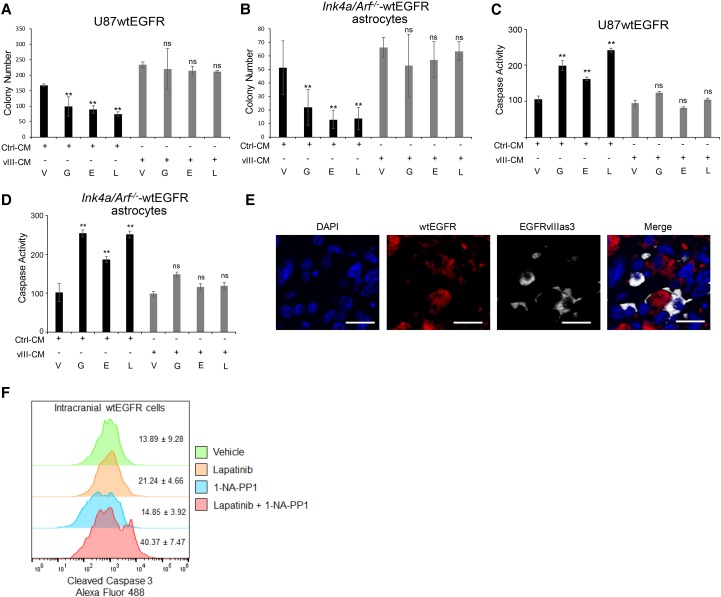
Figure 1.
EGFRvIII-secreted molecules exert anti-apoptotic action. (A,B) Soft agar colony formation assay quantification of U87wtEGFR cells treated with control medium obtained from parental U87MG cells (Ctrl-CM) or EGFRvIII cells (vIII-CM) in the presence of EGFR TKIs (A) and mAstr–Ink4/Arf−/−-wtEGFR cells treated with control medium obtained from parental mAstr–Ink4/Arf−/− cells or mAstr–Ink4/Arf−/−-vIII in the presence of EGFR TKIs (B). Colony number was determined from nine fields for each condition. (V) Vehicle; (G) gefitinib; (E) erlotinib; (L) lapatinib. (C,D) Caspase 3/7 activation assay of U87wtEGFR cells treated with control medium obtained from parental U87MG cells (Ctrl-CM) or EGFRvIII cells (vIII-CM) in the presence of EGFR TKIs (C) and mAstr-Ink4/Arf−/− wtEGFR cells treated with control medium obtained from parental mAstr–Ink4/Arf−/− cells (Ctrl-CM) or EGFRvIII cells (vIII-CM) in the presence of EGFR TKIs (D). Luminescence as relative light unit (RLU) intensity with blank subtracted and average values with standard deviations is shown. Percentage over control is reported. One-way ANOVA and two-tailed Student's t-test were used to compare samples. (**) P < 0.001; (ns) nonsignificant. (E) Intratumoral localization of EGFRvIIIas3 cells labeled with TurboFP635 (Alexa fluor 647) and U87wtEGFR cells labeled with iRFP720 (Alexa fluor 555). Bars, 20 µm. (F) Apoptotic wtEGFR cells in intracranial mixed tumors were identified by gating on EGFR antibody-stained cells (not shown) followed by detection of cleaved caspase 3. n = 4. Data are represented as mean ± SD.

