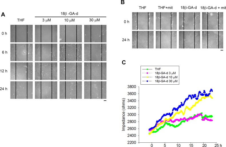
Fig 3. 18β-GA-d increased the migration of human skin fibroblasts.
(A) Human dermal fibroblasts were treated with 0.1% THF or different doses of 18β-GA-d (3, 10, and 30 μM) and observed at different times (6, 12, and 24 h). In vitro scratch wound healing assay was performed. Cell migration was analyzed using a phase-contrast microscope. Scale bar = 200 μm. (B) Human dermal fibroblasts were treated with mitomycin C (mit) (10 μg/ml) and 18β-GA-d (30 μM), and observed 24 h after treatment. Scale bar = 200 μm. (C) Human dermal fibroblasts on the wells with Electric Cell-substrate Impedance Sensing (ECIS) microelectrodes were wounded by a high electrical voltage. The medium was replaced with a new medium containing 0.1% THF or different doses of 18β-GA-d (3, 10, and 30 μM), and the cell migration was determined by ECIS. The migration of fibroblasts to the wounded area by electrodes was assayed using real-time measurement of the electrical impedance.

