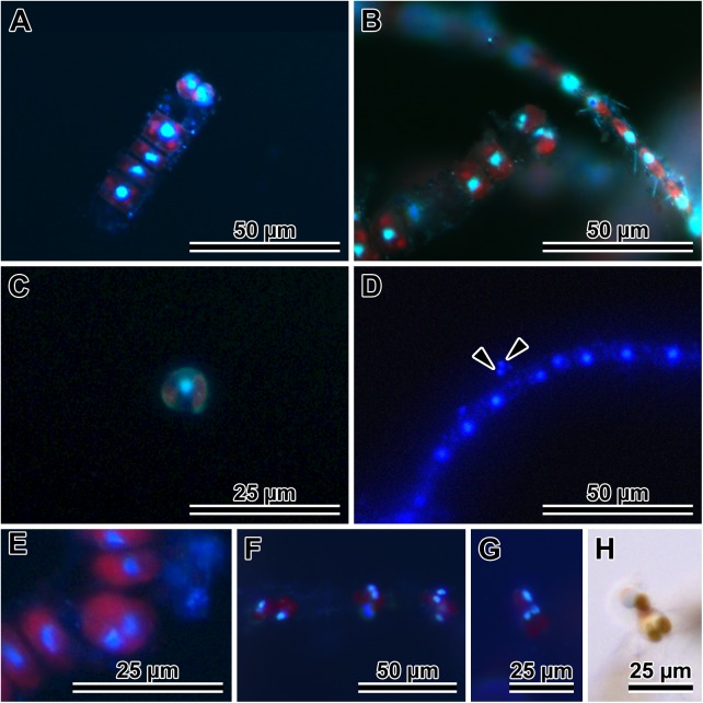
Fig 3. Gamete development and behaviour in P. tsawwassen.
(A and B) Stages in female gamete development. (A) A pair of uni-nucleate secondary oocytes. (B) Bi-nucleate cells destined female gametes, both characteristically perched on maternal valve copula. (C and D) Stages in male gamete development. (C) A uni-nucleate secondary spermatocyte liberated from paternal theca. (D) Bi-nucleate (arrowheads) male gamete free in mating dish environment. (E) Primary meiocyte after Meiosis I and before cytokinesis. (F) Three female gametangia with mostly bi-nucleate gametes perched on the thecae. (G) Gamete fusion, note bi-nucleated female gamete and uni-nucleate secondary spermatocyte in DAPI. (H) The same pair of live cells in LM; DAPI stained nuclei are blue, red is natural autofluorescence of chloroplasts.

