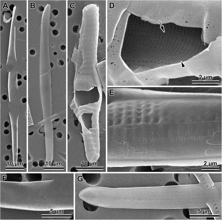
Fig 6. Initial valves.
(A) Internal and (B) external view of an initial epivalve of P. tsawwassen. (C) Partially damaged mature auxospore of P. staurophorum exposing the initial epivalve. (D) Enlarged section of the auxospore from C, showing initial epivalve (arrow) and one girdle band (arrowhead). (E) Another auxospore of P. staurophorum with initial hypovalve deposited internally to initial epitheca, two initial epicopulae cradle this hypovalve. (F) Close up of the internal surface of an initial epivalve from A, illustrating simple pores in mid-section. (G) External view of the perforation type near initial epivalve apex and on a copula of P. tsawwassen.

