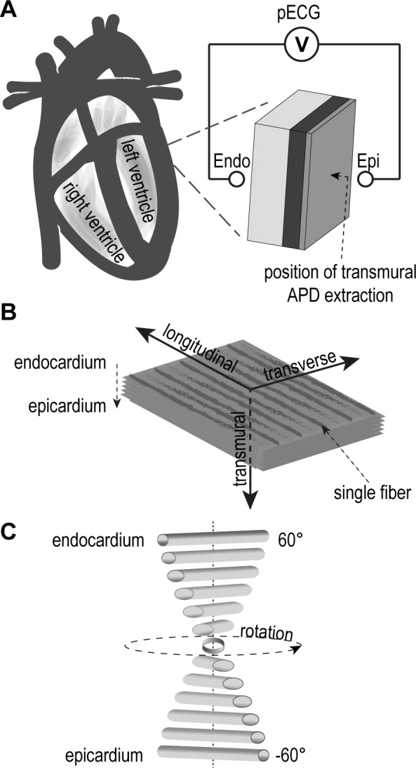
Fig 1. Schematic illustration of simulation setup.

(A) the extracted virtual ventricular wedge and the pECG setup, (B) the orientation in longitudinal (fiber direction of the sheet), transverse (parallel to fibers of the sheet) and transmural direction (from endocardium to epicardium) and (C) helical transmural fiber orientation.
