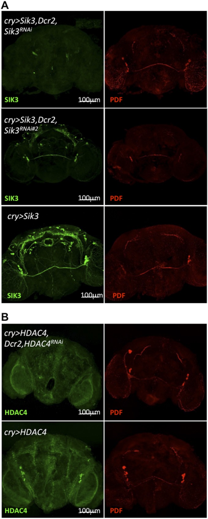
Fig. S4.
Sik3RNAi and HDAC4RNAi significantly reduce the expression of SIK3 and HDAC4 protein, respectively, in the brain. Epitope-tagged SIK3 (A), HDAC4 (B) and endogenous PDF were detected with anti-HA, anti-FLAG, and anti-PDF antisera, respectively. Reduction of SIK3 protein is much weaker in a second SIK3RNAi line (Sik3RNAi#2), the likely reason that Mz520 > Dcr2, Sik3RNAi#2 flies fail to show an MSDR phenotype (Table S2). Male brains were fixed at ZT5–8. This figure is related to Figs. 1 and 6.

