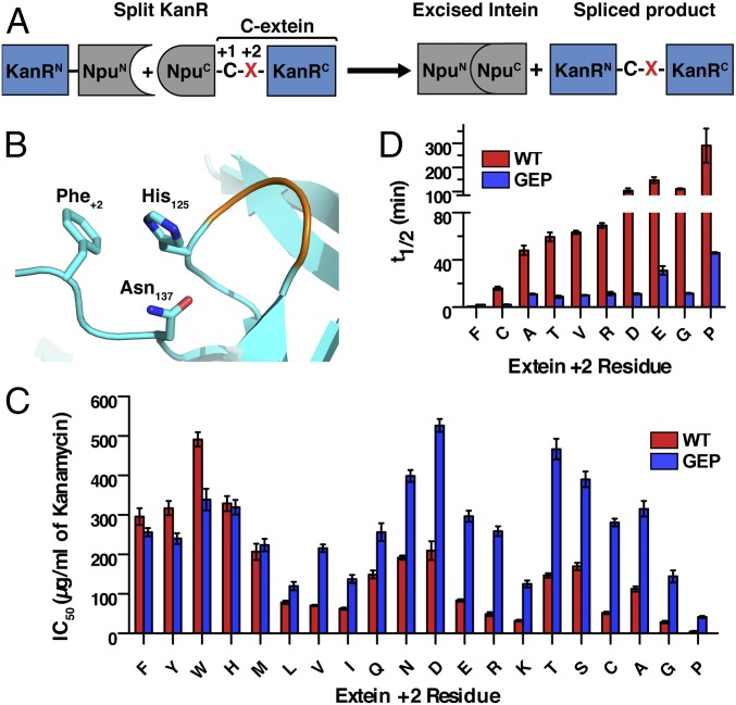
Fig. 2.
Engineering promiscuous splicing activity into the Npu DnaE split intein. (A) Schematic showing a PTS-dependent E. coli selection system. The kanamycin resistance protein, KanR, is split and fused to N- and C-intein fragments (NpuN and NpuC). The +2 C-extein residue (red X) can be varied in the system. (B) Depiction of the Npu active site (instantaneous structure from simulation; SI Appendix) highlighting the interaction among His125, Asn137, and Phe+2 (sticks), as well as the His125 loop (orange). (C) IC50 values for kanamycin resistance in E. coli for NpuWT (red) and NpuGEP (blue) across all +2 C-extein residues (mean ± SE, n = 3). (D) In vitro splicing half-lives of NpuWT (red) and NpuGEP (blue) with indicated +2 C-extein residues (mean ± SD, n = 3). Values for NpuWT Ala+2 and NpuWT Phe+2 are from previously reported data (10, 18).

