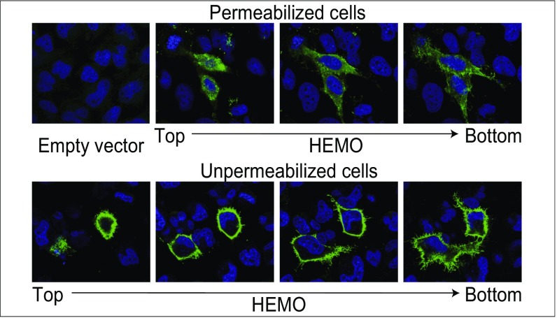
Fig. 2.
Immunofluorescence analysis of HEMO protein expression in transfected HeLa cells. Cells (HeLa) were transfected with the phCMV-HEMO expression vector (or an empty vector as a negative control), fixed, permeabilized (Upper) or not permeabilized (Lower), and stained for HEMO protein expression using a specific anti-HEMO polyclonal antibody (SI Methods). (Upper) Specific staining of the phCMV-HEMO transfected cells vs. empty vector transfected cells. (Lower) Successive confocal images show cell surface localization of the protein.

