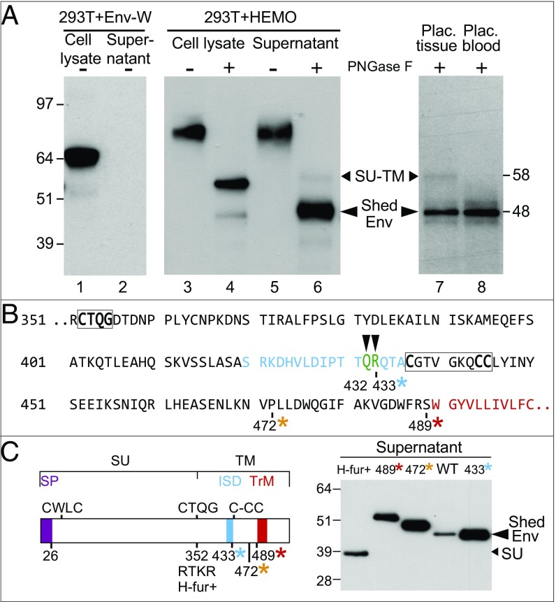
Fig. 3.
Characterization of the shed HEMO protein. (A) Detection of the shed HEMO Env protein by Western blot analysis. (Left) Detection of the syncytin-1 protein with the anti–Env-W polyclonal antibody (58) in the cell lysate of phCMV-Env-W transfected 293T cells. (Center and Right) Detection of the two forms of the HEMO protein (full-length SU-TM and Shed Env) with the anti-HEMO polyclonal antibody in the cell lysate and supernatant of phCMV-HEMO transfected 293T cells (Center) and first trimester placental tissue and placental blood (Right; matched representative samples from the same individual); samples were treated (+) or not (−) with PNGase F. (B) Detail of the shedding site amino acid sequence indicated by green capital letters. *Positions of the stop codons introduced in the mutants analyzed in C. (C) Migration pattern of the mutant HEMO forms analyzed as in A. (Left) Schematic representation of the HEMO protein with the same color code as in Fig. 1 and the stop codons of the generated mutants positioned together with that of the mutant with a reconstituted furin site (H-fur+; with an RTKR furin site). (Right) Supernatant of 293T cells transfected with the expression vectors for the WT and the mutant HEMO plasmids analyzed after PNGase F treatment, SDS gel electrophoresis, and Western blot as in A.

