Fig. 6.
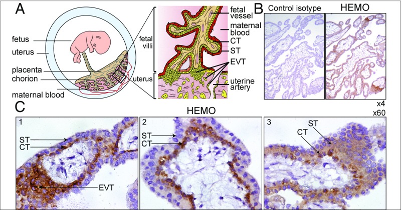
Immunohistochemical detection of the HEMO protein in first trimester human placenta. (A) Schematic representation of the fetoplacental unit with an enlarged anchored villus bathed by maternal blood and displaying the ST layer, the underlying mononucleated CT, and the invading EVT. Adapted from ref. 59. (B) Serial sections of multiple placental villi stained with a control IgG2a mouse isotype (Left) or the anti-HEMO mAb 2F7 (Right). (Magnification: 4×.) (C) Enlarged views of placental villi showing staining of CTs (1–3), with strong staining of EVTs (1) and diffuse staining of STs (3). (Magnification: 60×.)

