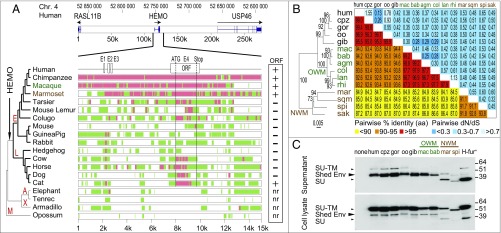
Fig. 9.
Sequence conservation and purifying selection of the HEMO gene in simians. (A) Syntenic conservation of the HEMO locus in mammalian species. The genomic locus of the HEMO gene on human chromosome 4 along with the surrounding RASL11B and USP46 genes (275 kb apart; genomic coordinates listed in Table S4) was recovered from the UCSC Genome Browser together with the syntenic loci of the indicated mammals from five major clades [Euarchontoglires (E), Laurasiatherians (L), Afrotherians (A), Xenarthres (X), and Marsupials M)]; exons and sense of transcription (arrows) are indicated. Exons of the HEMO gene (E1–E4) are shown on an enlarged view of the 15-kb HEMO locus together with the homology of the syntenic loci (analyzed using the MultiPipMaker alignment-building tool). Regions with significant homology as defined by the BLASTZ software (60) are shown as green boxes, and highly conserved regions (more than 100 bp without a gap displaying at least 70% identity) are shown as red boxes. Sequences with (+) or without (−) a full-length HEMO ORF are indicated on the right. nr, not relevant. (B) Purifying selection in simians. HEMO-based maximum likelihood phylogenetic tree was determined using nucleotide alignment of the HEMO genes (listed in Table S5 and Dataset S1). The horizontal branch length and scale indicate the percentage of nucleotide substitutions. Percentage bootstrap values obtained from 1,000 replicates are indicated at the nodes. Double-entry table for the pairwise percentage of amino acid sequence identity (lower triangle) and the pairwise value of dN/dS (upper triangle) between the HEMO gene from the various simian species listed on the phylogenetic tree to the left and listed in the same order in abbreviated form at the top. A color code is provided for both series of values. (C) Conservation of HEMO shedding in simians illustrated by Western blot analysis of 293T cells transfected with expression vectors for the indicated simian HEMO genes or the human HEMO mutant with a consensus furin site (H-fur+). Cell lysates and supernatants were harvested and treated with PNGase F before Western blot analysis with the polyclonal anti-HEMO antibody. The entire SU-TM HEMO protein is the main form observed in cell lysates, whereas the shed and the free SU form (for the NWM genes with a furin site and the H-fur+ mutant) are mainly observed in the supernatants. agm, African green monkey; bab, baboon; col, Angolan black-and-white colobus; cpz, chimpanzee; gib, gibbon; gor, gorilla; hum, human; lan, langur; mac, macaque; mar, marmoset; NWM, New World monkey; oo, orangutan; OWM, Old World monkey; rhi, golden snub-nosed monkey; sak, saki monkey; spi, spider monkey; sqm, squirrel monkey.

