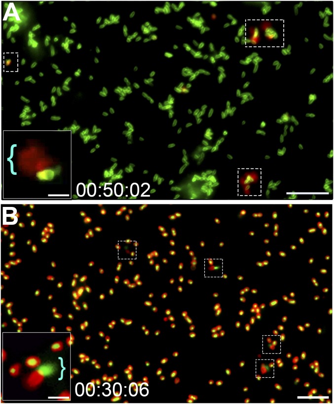
Fig. 2.
Subpopulation of NTHI released DNA from a single subpolar location along one long axis of the cell. (A) Time-lapse fluorescence video microscopy demonstrated DNA release from NTHI via a mechanism that did not involve development of large round cells or explosive cell lysis. Representative images show NTHI (green) cultured in medium that contained the membrane-impermeable dsDNA stain ethidium homodimer-2 (red), and thus a fluorescent red flare indicated DNA release from the bacterial cell (dashed boxes). Representative still frame was taken after 50 min of incubation. (Scale bar, 10 µm.) (A, Inset) Image of a cell with a red flare that indicated DNA release (blue bracket). (Scale bar, 2 μm.) (B) To validate that DNA was released from the bacterial cell, intracellular DNA was first stained with the cell-permeable stain Syto 9, which fluoresces green when bound to DNA, and the bacterial outer membrane was stained with FM4-64 (a red fluorescent membrane stain). Fluorescence time-lapse microscopy images confirmed that DNA was released from a subpolar location of a subset of cells. The presence of eDNA (now green) adjacent to a bacterial cell (now red) supported the proposed release mechanism (dashed boxes). (Scale bar, 10 μm.) (B, Inset) Magnified image of bacteria with a green flare that indicated DNA release (blue bracket). (Scale bar, 2 μm.)

