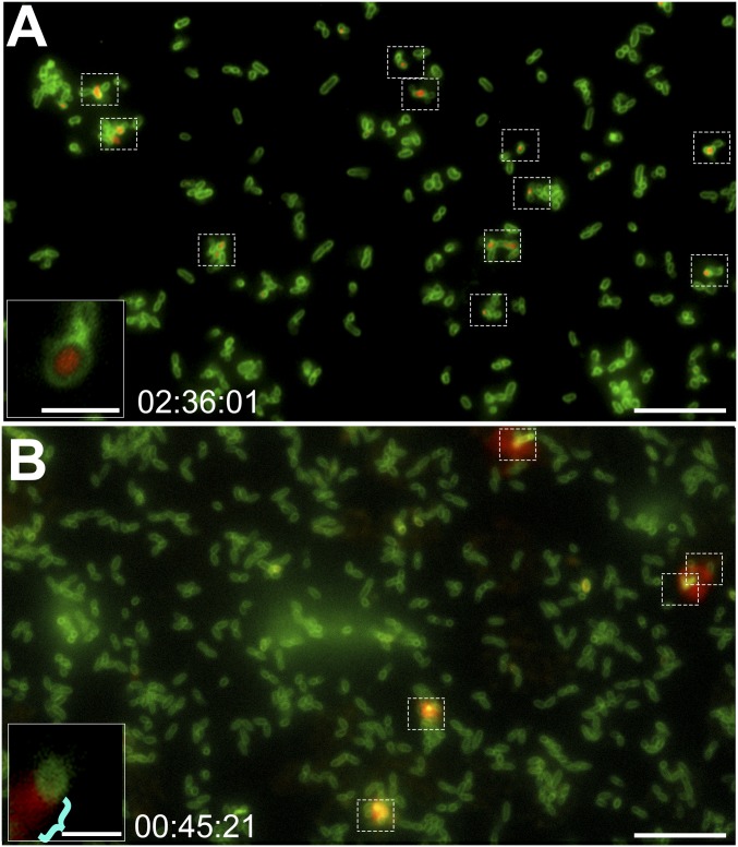
Fig. 6.
T4SS-like inner-membrane complex is required for the release of DNA from NTHI. Time-lapse fluorescence microscopy images of the ΔtraCG mutant and the complemented ΔtraCG mutant are shown. Time stamps indicate elapsed incubation time. (A) No DNA was released from the ΔtraCG mutant at any time point. However, a subset of cells (dashed boxes) had taken up the ethidium homodimer-2 DNA stain. (Scale bar, 10 µm.) (A, Inset) Magnified image of a ΔtraCG mutant cell showed a clear demarcation between red fluorescent DNA and green fluorescent outer membrane. (Scale bar, 2 μm.) (B) Complementation of the ΔtraCG mutant restored the characteristic DNA flare release phenotype (dashed boxes). (Scale bar, 10 µm.) (B, Inset) Magnified image of a complemented ΔtraCG mutant cell showed the characteristic red flare release phenotype (blue bracket). (Scale bar, 2 μm.)

