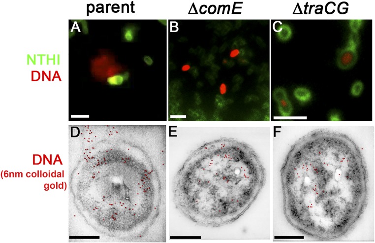
Fig. 7.
DNA was released from the parent but not from the ΔcomE or ΔtraCG mutants. (A–C) Duplicates of insets already presented in Figs. 2A, 5A, and 6A are repeated here for comparison (please see respective Fig. 2 legends for a detailed description). (D–F) ImmunoSTEM for the presence of DNA-confirmed fluorescent labeling seen in A–C. (D) A subset of NTHI parent cells positively labeled for DNA within the cytoplasm, periplasm, and extracellular environment as seen by 6-nm gold particles (pseudocolored red based on detection of secondary electrons; see Fig. S1 for additional images). (E) Subset of ΔcomE mutant cells positively labeled for DNA within the cytoplasm and in the periplasm, but no extracellular labeling was detected. (F) DNA was detected within the cytoplasm of the ΔtraCG mutant cells with no labeling within either the periplasm or extracellular space. (Scale bars, 200 nm.)

