Fig. 8.
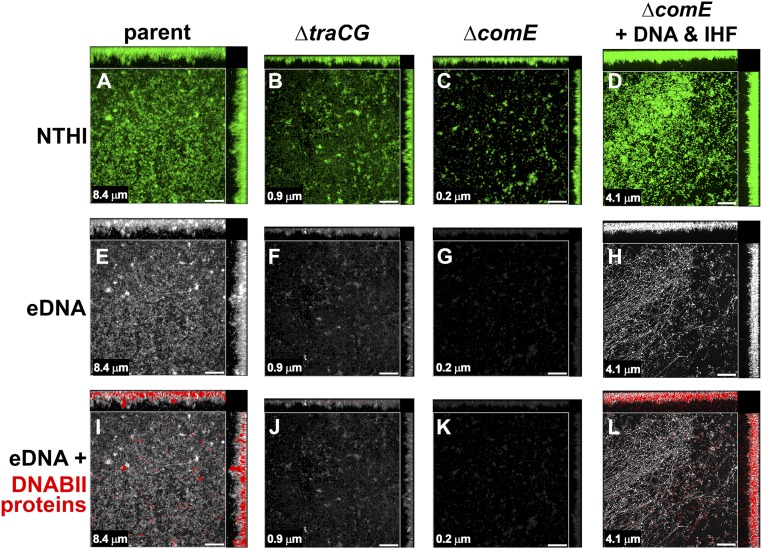
eDNA and DNABII proteins were present in biofilms formed by the parental isolate but not the ΔcomE or ΔtraCG mutants. Biofilms were immunolabeled for DNA and DNABII proteins. (A–C) Bacteria within the biofilms were stained with FM1-43 outer-membrane stain (green fluorescence) with both top-down and orthogonal views of representative biofilms shown. (E–G) eDNA (white) is visible throughout the biofilm formed by the parental isolate with minimal labeling in biofilms formed by either mutant. (I–K) DNABII proteins (red) were visualized in addition to eDNA (white) throughout biofilms formed by the parental isolate. In comparison, minimal to no labeling for DNABII proteins (red) was observed in biofilms formed by either of the mutants. Addition of exogenous NTHI DNA and a DNABII protein (IHF) restored ΔcomE biofilm characteristics comparable to the parent (compare D, H, and L with A, E, and I). (Scale bars, 20 μm.) Note: Mean biofilm height as determined by COMSTAT2 analysis is indicated in the lower left-hand corner of each panel.

