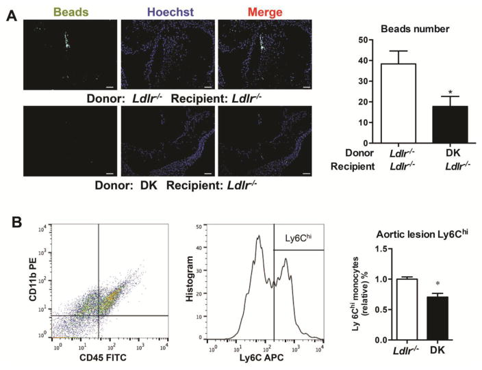
Figure 4. sEH deficiency decreased infiltration of Ly6Chi subset of infiltration in aortic root.
Mice with bone marrow transplantation were injected intravenously with 250 ml clodronate-containing liposomes for 2 days, then mice were injected with Fluoresbrite Plain YG microspheres (Polysciences), 1 mm in diameter, to label the newly-generated Ly6Chi monocyte subset. After a 6-week WTD, aortas of Ldlr−/− female mice were excised and invading beads in aorta sections were quantified. (A) Representative fluorescence-labeled Ly6Chi monocytes in aortic roots. Scale bars, 100μm. Quantification of bead number is on the right. n≥10, (B) The isolated cells of aorta from Ldlr−/− and DK mice fed with 6-week WTD were digested and stained by CD45, CD11b and Ly6C. The CD45+CD11b+Ly6Chi cells were quantified. n=7. Data are mean ±SEM. *p<0.05.

