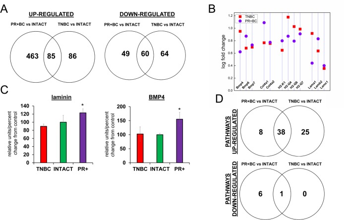
Figure 1. Next generation sequencing‐based analysis of gene expression in the PFC tissues of intact and TNBC and PR+Bcbearing TumorGraft mice.
(A) Venn diagram showing genes that were significantly different between TNBC and PR+BC mice, as compared to intact controls; (B) Fold changes in the levels of expression of selected genes; (C) Western immunoblotting analysis of laminin and BMP4 proteins in the PFC tissues of TNBC and PR+BC mice; data are shown as relative units/percent change of control. Due to size difference the same membrane was used for both proteins. * p<0.05, Student's t‐test; (D) Summary of molecular pathways that were altered in the PFCs of TNBC and PR+BC mice as compared to intact controls. The Pathview/KEGG analysis was used to determine differentially affected pathways.

